Figure 2 ~ 10과 Table 1~ 4의 결과와 같이, 샘플인 세가지 종류의 Peptide들은 폭넓은 범위에서 우수한 감도와
재현성을 보여준다.
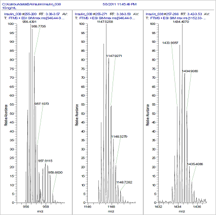
Figure 2. Spectra of the acquired mass peaks of human insulin
at 70,000 resolution.
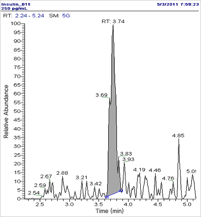
Figure 3. Chromatogram of 250 pg.mL-1 human insulin (LLOQ).
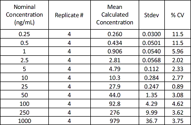
Table 1. Insulin summary table; dynamic range
from 250 pg.mL.-1 to 1000 ng.mL.-1
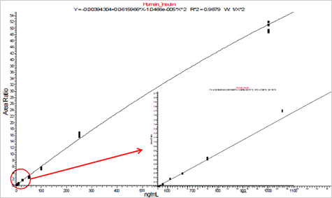
Figure 4. Insulin calibration curve, dynamic range
from 250 pg.mL.m-1 to 1000 ng.mL.-1
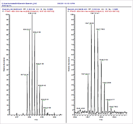
Figure 5. Spectra of the acquired mass peaks
of Exendin-4 at 70,000 resolution.
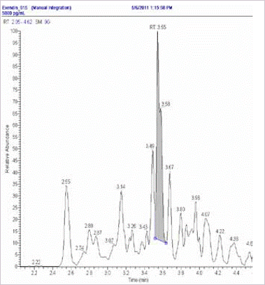
Figure 6. Chromatogram of 5 ng.mL-1 Exendin-4 (LLOQ).
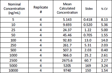
Table 2. Exendin-4 calibration table; dynamic range
from 5 ng.mL.-1 to 10000 ng.mL.-1
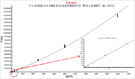
Figure 7. Exendin-4 calibration curve, dynamic range
from 5 ng.mL.-1 to 10 μg.mL.-1
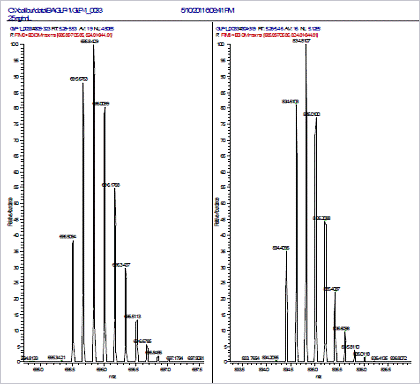
Figure 8. Spectra of the acquired mass peaks of GLP-1
at 70,000 resolution.
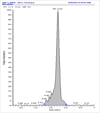
Figure 9. Chromatogram of 5 ng.mL-1 GLP-1 (LLOQ).
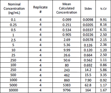
Table 3. GLP-1 summary table; dynamic range
from 0.1 ng.mL.-1 to 10000 ng.mL.-1
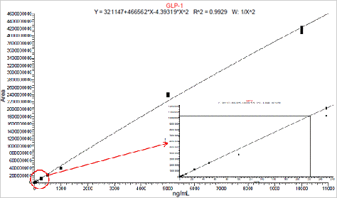
Figure 10. GLP-1 calibration curve; dynamic range
from 5 ng.mL.-1 to 10 μg.mL.-1
|